Diabetic foot represents a complication of diabetes that can lead to foot ulcers and other serious conditions. 3D Finite Element (FE) analysis enables dynamic characterisation of different loads within the foot.
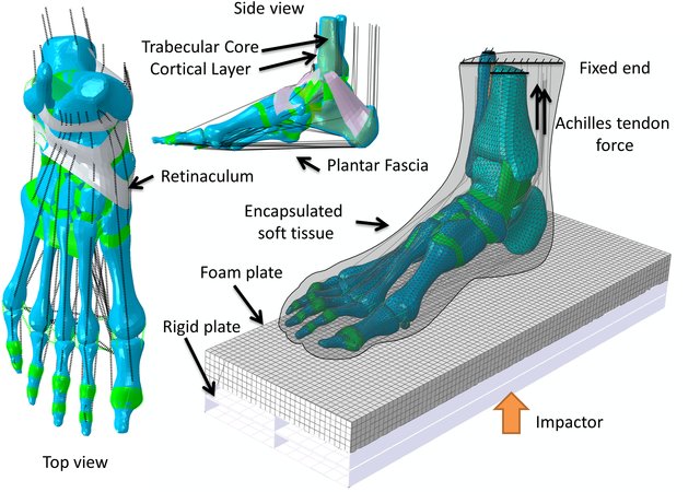
Annamaria Guiotto from University of Padova, Italy developed a subject-specific 3D FE model of a diabetic neuropathic and a healthy subject’s foot from MRI data. Experimental analysis of the biomechanics of the foot was combined with FE simulations of foot models generated in Simpleware software. Subject-specific kinematics and kinetics data acquired during gait analysis were used in the simulations.
IMAGE PROCESSING
Experimental analysis of the foot was conducted on ten healthy and ten diabetic neuropathic subjects, and hindfoot, midfoot, forefoot, subsegments and tibia 3D kinematics were estimated together with 3D foot subsegments kinetics and plantar pressure (PP). The geometries of the healthy and diabetic feet were then obtained using an MRI scanner (Philips Achieva; Siemens Avanto).
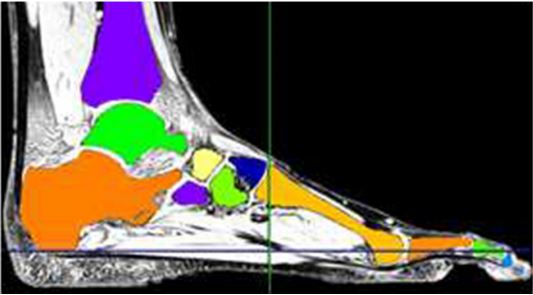
Data was loaded in Simpleware ScanIP and segmented into 30 bones, cartilage and skin, generating a 3D model of the whole foot and ankle. The phalanges of the foot were separated from the metatarsals, and the plantar fascia, Achilles tendon and short plantar ligaments were inserted within the model.
MESHING
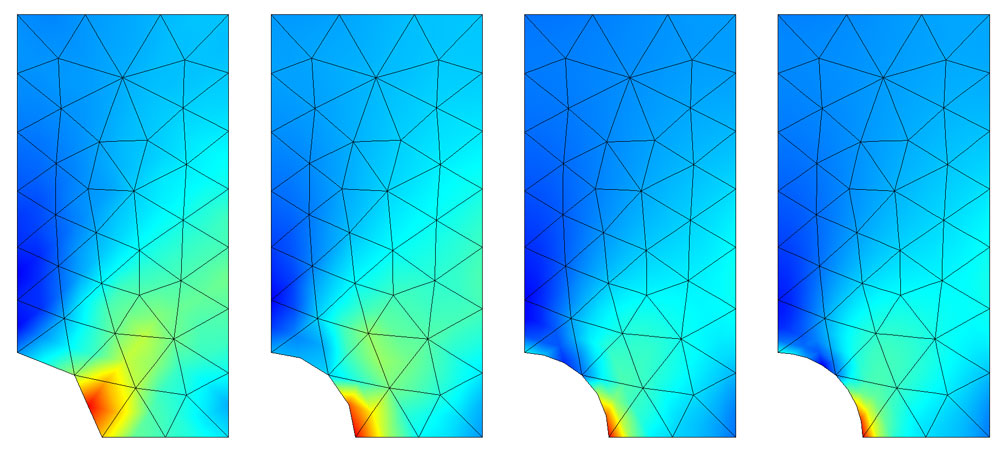
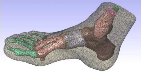
A FE mesh was generated in Simpleware +FE using tetrahedral elements. The mesh included conforming interfaces, resulting in no gaps or overlaps between parts of the model. The +FE Free algorithm was used with a minimum and maximum edge length of 6/8 mm and a 0.4 target maximum error. The final mesh was exported directly to Abaqus.
A horizontal rectangular element was drawn in Abaqus under the foot to simulate ground support. Material properties were adapted from the literature, and the diabetic foot modelled with increased stiffness values for the soft tissues. Four different loading conditions were applied considering different phases of the stance phase of gait (heel strike, loading response, midstance and push off), and run with the prepared kinematics and kinetics data.
RESULTS and CONCLUSIONS
FE simulations were run with the kinematics and kinetics data of the healthy and diabetic neuropathic FE models. Validation of the models was carried out by comparing experimental plantar pressures (PP) and the simulated ones (peak and mean values in the 3-foot subareas and in the whole foot).
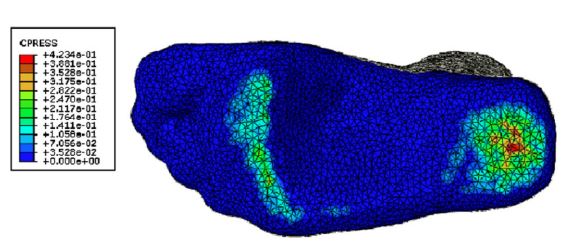
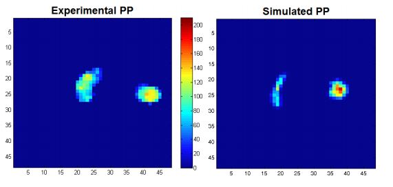
A good agreement was found between the predicted and measured PP distribution when the kinematic kinetic data of the healthy and diabetic neuropathic feet were used. However, when gait data from the two groups was adapted for the simulations, the healthy foot underestimated the PP and overestimated contact surfaces. The neuropathic group results were in better agreement.
In general, comparison of experimental and simulated results demonstrated the effectiveness of subject-specific FE models for obtaining reliable simulated PP during gait. This knowledge is crucial in understanding the aetiology of the diabetic foot.
Guiotto, A. et. al. (2014). Journal of Biomechanics. 47(12):3064-3071.